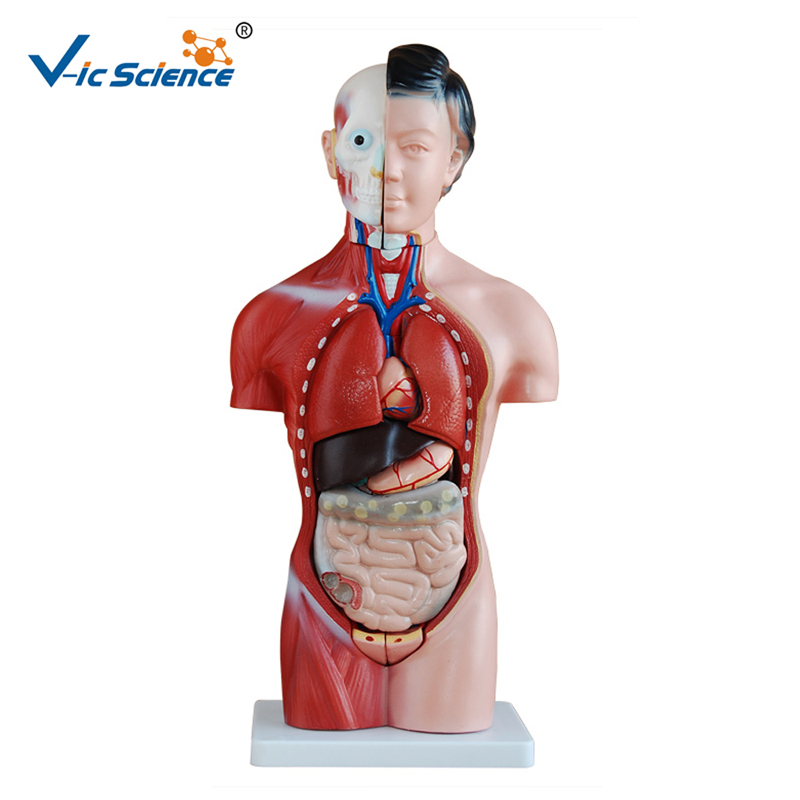
Human trunk model shows the normal position, morphological structure and mutual relationship of human organs. It is a model applied to human anatomy teaching. It can enable students to understand the relationship between adult normal posture and internal organs, and show the position and structure of main organs. It is widely used in many fields such as medical affairs and biology.
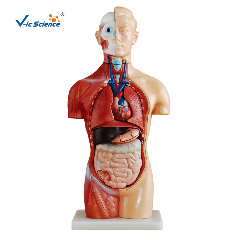
It shows the position of human internal organs and the anatomical shape and structure of the head, showing three systems: respiration, digestion and urinary system.
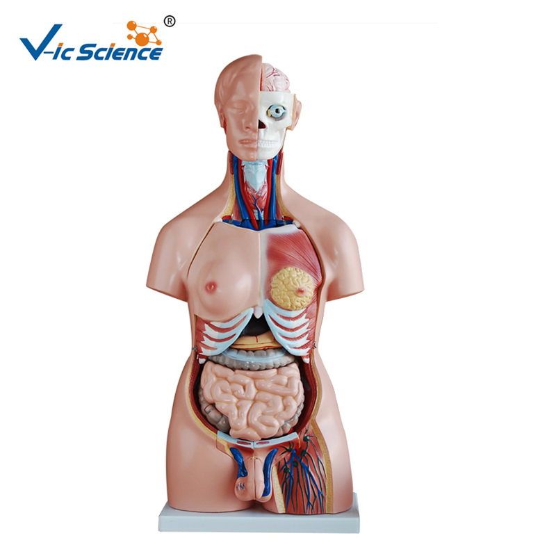
The right half of the head and neck shows the skull, masseter muscle, temporal muscle and other structures. There are eyeballs in the orbit. The sagittal section is made at the head and neck. The skull cavity contains the right hemisphere of the brain. There are 12 pairs of brain nerves on the ventral side of the brain, showing the nasal cavity, oral cavity, throat, laryngeal cavity, laryngeal chamber, glottic fissure and parathyroid gland behind the lateral lobe of the thyroid gland.
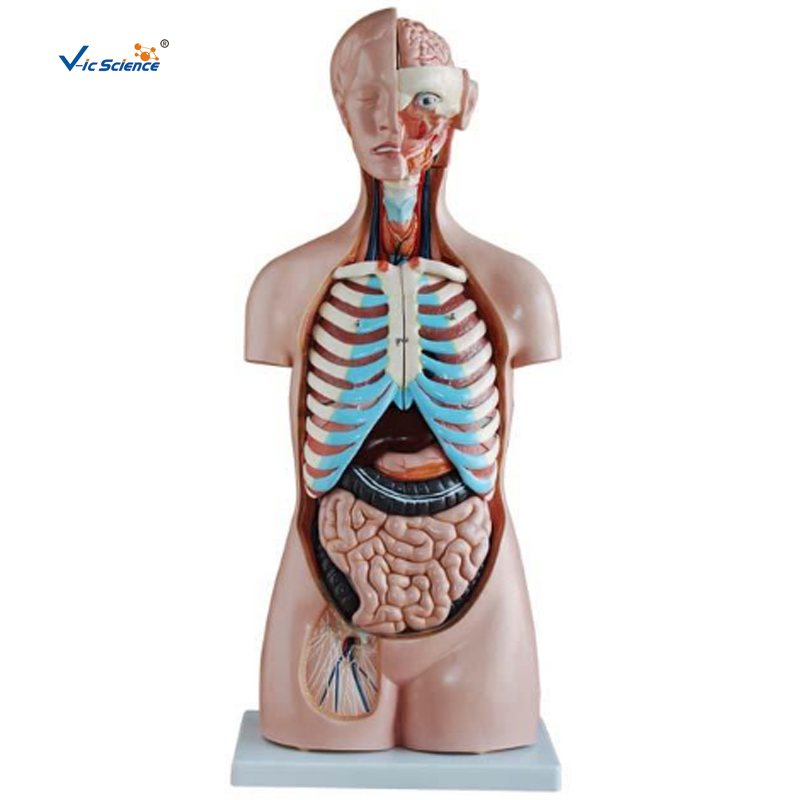
The frontal sections of the two lungs in the thoracic cavity show the structure of the lungs. The coronary anatomy of the heart shows the structural similarities and differences between the left and right atrioventricular. The cardiac vessels include superior and inferior vena cava, pulmonary arteriovenous and aorta for explaining the application of large and small blood circulation..
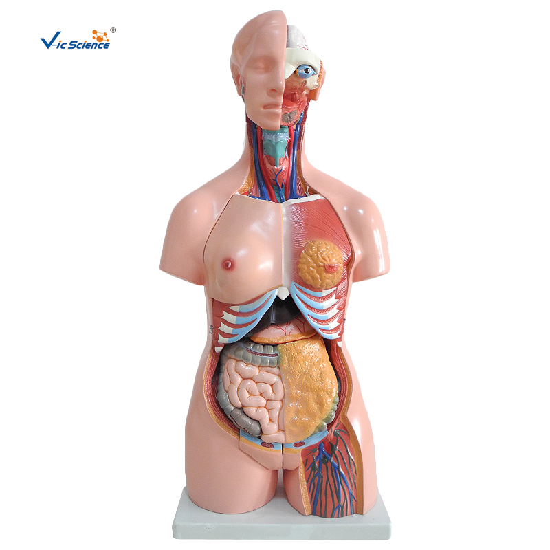
Below the septum is the abdominal cavity, containing liver, stomach, pancreas, spleen, kidney, bladder and other internal organs. The anatomy shows the structure of digestive tube wall such as descending duodenum, cecum and small jejunum. The anatomical surface of the right kidney shows the structure of cortex, medulla and renal pelvis.