1. Take materials
The fresher the tissue, the better. Generally, human tissue is in vitro, and animal tissue is quickly fixed after death to ensure the original morphological structure.
2. Fixing
The most commonly used fixatives are 10% formaldehyde fixative and 95% ethanol fixative.
3. Dehydrated transparent
After the specimen is fixed and washed, the tissue contains more water, and the water in the tissue block must be replaced. This process is called dehydration. Whether paraffin section or collodion section is used, the water contained in the tissue must be removed. Because the aqueous tissue is incompatible with paraffin, collodion and other embedding materials, the commonly used dehydrating agent is a series of ethanol with different concentrations.
4. Wax dipping and embedding
(1) The paraffin is immersed in the tissue to replace the transparent agent contained in the tissue.
(2) Embedding: put the tissue soaked in wax into the melted solid paraffin. After the paraffin solidifies, the tissue is wrapped in it, which is called wax block. This process is called embedding.
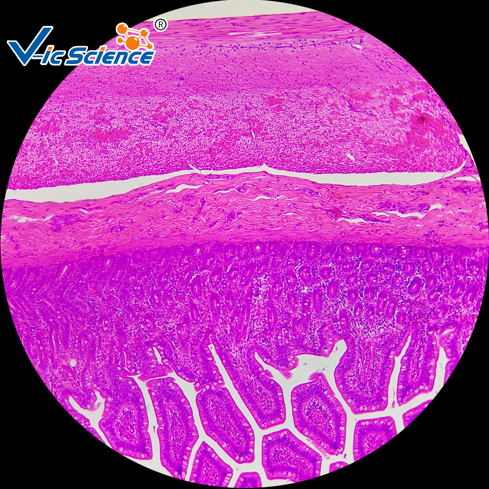
5. Slice and patch
The thickness regulator of the slicer is engraved with 0 ~ 50 μ M or 0 ~ 25 μ m. Its thickness can be arbitrarily selected, and the thickness of paraffin section is generally 4 ~ 6 μ m.
Patch and baking: after the slice is fully flattened on the constant temperature water surface, take the wax slice to the middle section of the slide, pour out the residual water on the slide, put it into the 60 ~ 65 ℃ incubator or the oven of the slice bleaching and baking temperature controller, bake the slice for 15 ~ 30 minutes, and remove the paraffin in the dissolved tissue gap.
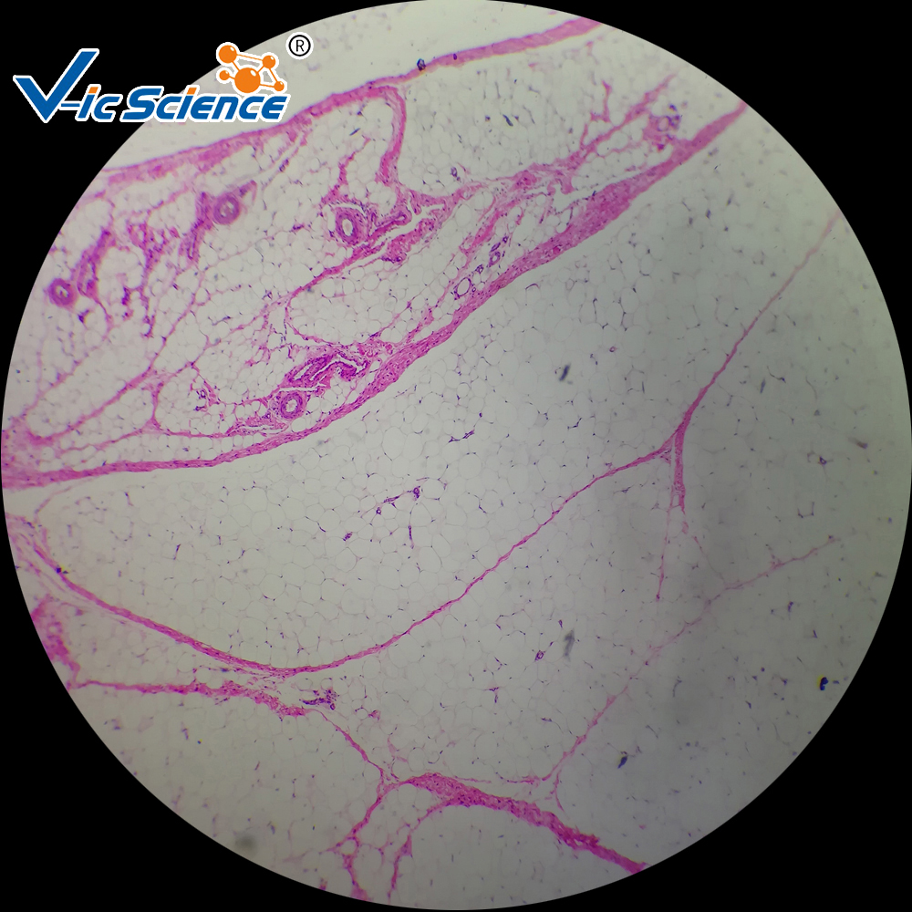
6. Dyeing and sealing