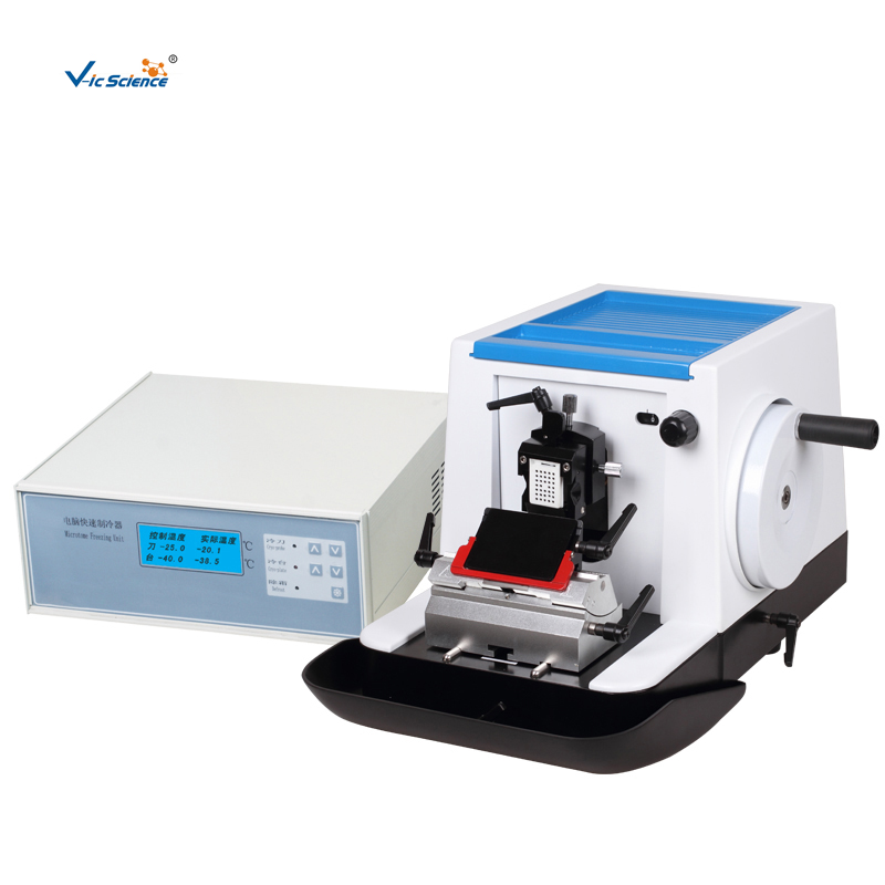
The most widely used method: paraffin section is not only used to observe the morphological structure of normal cell tissue, but also the main method for pathology and forensic science to study, observe and judge the morphological changes of cell tissue, but also has been widely used in many other scientific fields.
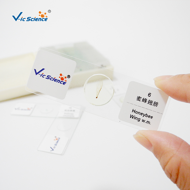
In teaching, most of the specimens observed under light microscope are prepared by paraffin section method. Most living cells or tissues are colorless and transparent, and there is a lack of contrast between various tissues and various structures in cells, which is not easy to be clearly distinguished under general light microscope; After leaving the body, the tissue will soon die, become corrupt and lose its original normal structure.Therefore, the tissue should be fixed, embedded in paraffin, sectioned and stained to avoid cell death, and its morphological structure can be clearly identified.
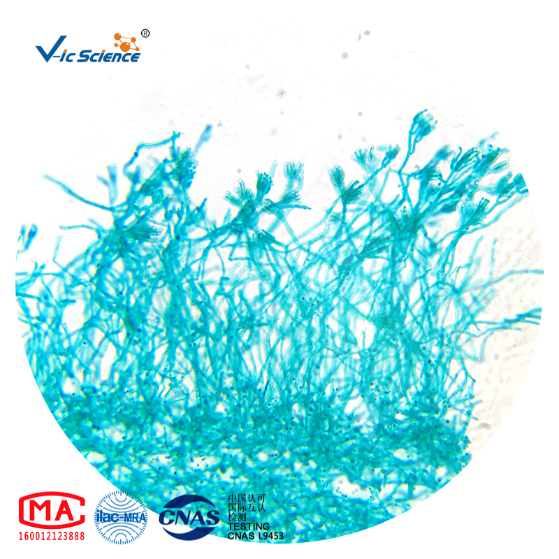
The basic technology of paraffin section production paraffin section method includes the steps of material extraction, fixation, washing and dehydration, transparency, wax immersion, embedding, slicing and sticking, dewaxing, dyeing, dehydration, transparency, sealing, etc. It takes several days for the general tissue to be fixed from the material to the seal to make the slide specimen, but the specimen can be preserved for a long time, which is a permanent microscopic slide specimen.